
Welcome to an informative look at one of the simplest yet most transformative tools in modern dental practice: the intraoral camera. At Back Bay Dental, serving the greater Great Lakes and Lake Winnipesaukee region in Wolfeboro, NH, we believe that education is key to empowering our community to make informed decisions about their health. The intraoral camera is a tiny, pen-sized device that captures high-resolution, full-color images and video inside the mouth. For decades, a dentist’s primary diagnostic tools relied heavily on small mirrors, tactile explorers, and two-dimensional X-rays. While these traditional tools remain fundamental, they often limit the patient’s ability to see and understand what the practitioner was truly describing. This gap between the clinical assessment and the patient’s comprehension often hinders collaborative decision-making.
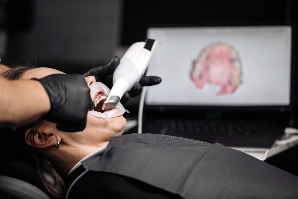
The introduction of the intraoral camera bridges this gap by providing an immediate, clear, and magnified view of the patient’s oral condition. The small camera is gently guided around the mouth, illuminating and capturing images of teeth, gums, and other oral tissues. These live images are instantly projected onto a chairside monitor, turning the abstract descriptions of a diagnosis into a concrete, visible reality. This shift from purely verbal explanation to vivid visual evidence marks a significant advancement in patient communication and clinical accuracy. The capability to visually document the starting point of any issue, from minor cracks to early-stage gum disease, allows for meticulous record-keeping and a powerful educational platform.
Enhanced Diagnostic Precision and Early Detection
The high magnification and intense illumination provided by the intraoral camera significantly enhance diagnostic precision, allowing dental professionals to identify issues that might be missed during a standard visual and tactile examination. The camera can zoom in hundreds of times, bringing minute details of the tooth structure into sharp focus. This is particularly valuable for detecting tiny problems, such as hairline fractures, the earliest stages of decay (caries) that haven’t yet shown up on an X-ray, or the subtle signs of inflammation along the gum line. Catching these problems early is crucial, as early intervention is often less invasive, less costly, and more effective than treating advanced disease.
Furthermore, the lighting capabilities of the intraoral camera are vastly superior to the ambient lighting in the operatory or even a focused overhead lamp. This intense, targeted light can reveal discrepancies in tooth shade, detect marginal breakdown around existing fillings or crowns, and illuminate the deep fissures and grooves on the chewing surfaces of back teeth where bacteria often hide. The ability to freeze and save these images creates a valuable photographic record that can be compared over time to monitor conditions like excessive wear, recession of the gums, or changes in suspicious lesions. By using this tool, the practitioner can move beyond subjective assessment to a documented, evidence-based diagnosis, leading to a more reliable and complete treatment plan.
Revolutionizing Patient Education and Trust
One of the most profound benefits of the intraoral camera is its power to revolutionize patient education and build trust in the diagnosis and recommended treatment. Before this technology, a dentist might point to an area in the mouth and use technical terminology to explain a problem, such as a “recurrent carious lesion on the mesial margin of the amalgam restoration.” For many patients, this description remains abstract, leaving them feeling disconnected from their diagnosis and potentially hesitant to proceed with treatment they don’t fully understand.
With the intraoral camera, the dentist can show the patient a crystal-clear, magnified image of the exact area of concern right on the monitor. Seeing a dark spot of decay, a chipped tooth, or an inflamed gum line eliminates guesswork and allows the patient to become an active participant in their care. The visual evidence serves as a powerful teaching tool, making the need for treatment tangible and urgent. When patients can literally see the proof of their condition, they gain a greater understanding of why a procedure is necessary, thereby increasing their acceptance of recommended care. This transparency, fostered by shared visual information, leads to stronger patient-professional trust, improving compliance with both treatment plans and long-term preventive care strategies.
Enhanced Documentation and Insurance Support
Beyond the immediate diagnostic and educational benefits, the intraoral camera provides superior documentation that is critical for comprehensive record-keeping and is immensely helpful when communicating with insurance providers. Every image captured can be stored digitally in the patient’s file, creating a comprehensive, organized visual history of their oral health over many years. This chronological record of photographs serves as an essential benchmark for tracking the progression or stability of various conditions. For instance, an early-stage crack that is placed under observation can be reliably monitored with annual intraoral photographs, ensuring that treatment is initiated only when necessary.
Furthermore, these high-quality, clear images are often invaluable when submitting claims to dental insurance companies. Historically, insurance providers might delay or deny treatment authorization based solely on written documentation or traditional X-rays, which may not fully illustrate the severity or nature of a problem. A clear, magnified photograph showing a failing filling, a visible fracture, or significant soft tissue inflammation provides incontrovertible visual evidence of the need for the procedure. This robust visual documentation streamlines the pre-authorization process, reduces claim disputes, and helps ensure that patients receive timely approval for the care they need, removing a potential barrier to essential treatment.
- Key Advantages for Comprehensive Care:
- Provides visual proof for insurance claim approval.
- Allows for accurate monitoring of non-urgent conditions over time.
- Creates a complete digital record for specialist referrals and continuity of care.
- Offers immediate verification of the finished restoration placement.
Improved Communication and Treatment Planning
The intraoral camera plays a critical role in improving communication during collaborative treatment planning, particularly when multiple professionals are involved in a patient’s care. When a general practitioner needs to refer a patient to a specialist—such as an endodontist, periodontist, or oral surgeon—the ability to digitally share a high-resolution image of the specific area of concern facilitates a much smoother handoff. Instead of relying solely on written notes and X-rays, the specialist can immediately see the clinical presentation of the issue, which aids in pre-consultation diagnosis and preparation.
For the patient, this technology enhances the treatment planning phase by allowing them to preview their mouth before and after potential treatment options. For example, suppose a dentist is discussing options for cosmetic improvements. In that case, the intraoral camera can capture the existing condition, and software may allow for a digital mock-up or smile design to be visually superimposed, giving the patient a realistic expectation of the final result. This clear communication throughout the planning phase—guided by undeniable visual evidence—ensures that the patient’s goals and the clinical reality are perfectly aligned, leading to higher satisfaction and more predictable outcomes for Dr. Nicholas Ciancarelli and any specialists involved.
Conclusion
The integration of the intraoral camera into the daily practice of dentistry has fundamentally shifted the diagnostic and educational landscape. By offering high-resolution, magnified visuals, it fosters a level of precision, transparency, and patient engagement that traditional methods simply cannot match. For individuals in the greater Great Lakes and Lake Winnipesaukee region, embracing such advanced tools means receiving more accurate diagnoses, understanding treatment needs more clearly, and participating proactively in long-term oral health management. Thank you for learning about this powerful tool with Back Bay Dental.
Resources
American Dental Association. (2023). Dental Technology and Modern Practice. ADA Publishing.
Schuller, M. (2018). Intraoral Imaging: The Complete Guide to Mastering the Intraoral Camera. Dentistry Today.
Timmerman, A. E., Van der Sanden, M. A. W. C. M., & Van der Weijden, F. A. (2018). The Influence of Intra-Oral Cameras on Patient-Reported Satisfaction and Treatment Acceptance: A Systematic Review. Journal of Dentistry.