
In recent years, dental care has witnessed remarkable technological progress—one of the most transformative being the evolution of x-ray imaging. Traditional film x-rays, once the gold standard, are rapidly being replaced by more advanced digital x-ray systems that provide better imaging, improved diagnostics, and increased patient safety. At Back Bay Dental in Wolfeboro, NH, serving Carroll County and the greater Lake Winnipesaukee region, we believe in keeping patients informed about the technology shaping the future of dentistry. In this blog, we’ll explore how digital x-ray advancements are improving oral health care for both patients and providers alike.
From Film to Digital: A Technological Leap
Traditional dental x-rays required film processing in darkrooms, with chemicals, delays, and the inherent risk of human error. These analog systems produced quality images, but the process was time-consuming and not environmentally friendly. Digital x-rays revolutionized this process by allowing images to be captured electronically and viewed instantly on a computer screen. Instead of waiting for film to develop, dental professionals can now analyze high-resolution images within seconds.
Digital x-ray systems use electronic sensors rather than traditional photographic film. This enables immediate visualization and easy enhancement of images. Dentists can zoom in, adjust contrast, and apply filters to highlight problem areas more clearly—something that simply wasn’t possible with film-based systems. This improved clarity can assist in identifying issues like small cavities, early signs of bone loss, or hairline fractures that may have gone unnoticed in the past. The switch from film to digital represents more than just convenience—it’s a leap forward in diagnostic precision.
Lower Radiation Exposure for Patients
One of the most significant health-related benefits of digital x-ray technology is its reduction in radiation exposure. Traditional x-ray systems, while generally safe, still exposed patients to more radiation than modern digital systems. Digital radiography requires significantly less radiation—up to 90% less in some cases—making it a much safer alternative, especially for patients who require frequent imaging.
Radiation safety is an important consideration in dental care, particularly for children, pregnant individuals, and patients undergoing complex treatment plans. Because digital systems capture images more efficiently and require fewer retakes, the cumulative exposure over time is drastically reduced. Patients can feel more confident knowing their oral health is being assessed with technology that prioritizes safety without compromising diagnostic effectiveness.
Enhanced Diagnostic Capabilities
The higher resolution and adjustability of digital x-rays allow for earlier and more accurate detection of dental problems. When dentists can detect conditions like decay, infections, or bone changes earlier, it often leads to less invasive treatment options and better outcomes. In complex cases—such as orthodontic assessments, implant planning, or evaluating jaw joint disorders—digital x-rays provide detailed images that support more precise treatment planning.
Digital radiography also supports integration with advanced imaging software, such as 3D modeling and computer-aided design (CAD) systems. This means a digital x-ray can be the first step in designing crowns, bridges, or aligners that are custom-fit to the patient’s unique anatomy. For surgical procedures, 3D cone beam imaging (CBCT) gives a detailed view of bone structure, nerves, and sinuses—allowing for better surgical outcomes and reduced risk of complications. These advancements are helping clinicians move beyond basic diagnosis into highly personalized, digitally guided care.
Environmental and Practical Benefits
Digital x-ray systems don’t just benefit patients—they’re also a more environmentally responsible choice. Traditional x-ray development relies on chemical processing, including hazardous substances that must be disposed of carefully to avoid environmental contamination. Digital imaging eliminates the need for these chemicals entirely, creating a cleaner and greener practice overall.
In addition, digital x-rays support more efficient dental practices by streamlining data storage and communication. Images can be easily stored in electronic health records and instantly shared with specialists, insurance providers, or other members of a patient’s care team. This level of integration reduces wait times for referrals and promotes better interdisciplinary care. Digital records also make it easier to track changes in a patient’s oral health over time, supporting more proactive and long-term treatment planning.
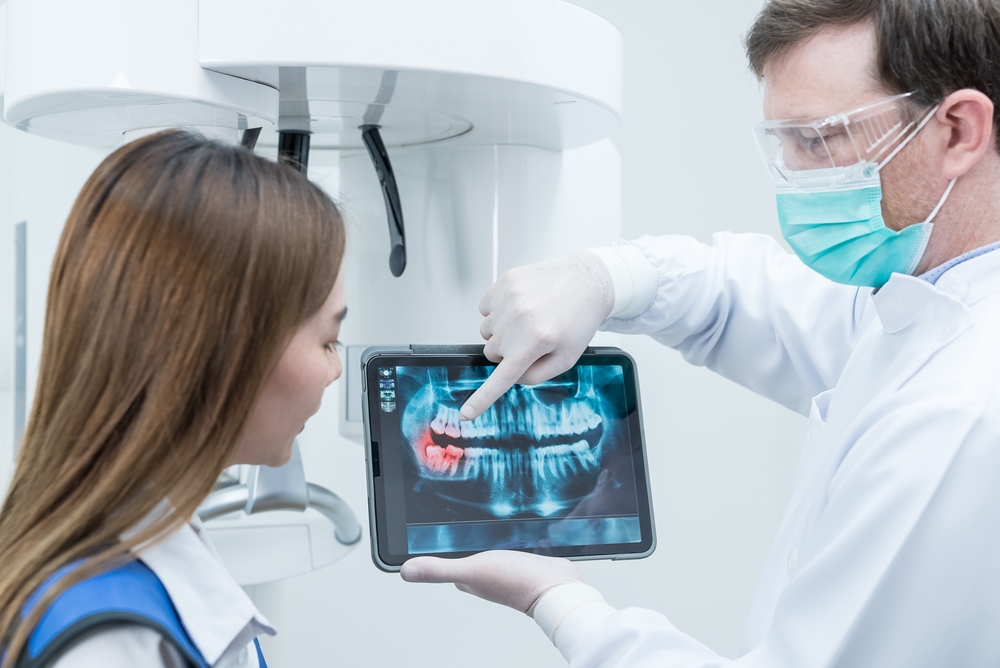
Patient Education and Engagement
One of the often-overlooked benefits of digital x-rays is how they improve patient communication and understanding. When patients can see their x-rays clearly on a screen and have their dentist explain findings in real time, it helps build trust and encourages more active participation in treatment decisions. For many patients, visualizing the problem can make a significant difference in understanding the importance of recommended treatments or preventive care.
Digital x-rays can be displayed alongside other diagnostic tools, such as intraoral camera images, to provide a comprehensive view of the patient’s oral health. This helps demystify dental terminology and turns what could be an abstract conversation into a concrete, visual discussion. Increased transparency supports stronger dentist-patient relationships and empowers individuals to take control of their oral health journey.
Looking Ahead: The Continuing Evolution of Dental Imaging
Digital x-ray technology continues to evolve, with future advancements likely to focus on even faster imaging speeds, higher resolutions, and greater integration with artificial intelligence (AI). AI-assisted diagnostics are already being explored in some practices, with software that can flag potential issues or track subtle changes over time. These tools may help standardize diagnosis and reduce human error, making dental care even more consistent and reliable.
As imaging continues to advance, it’s clear that digital x-ray technology is not just a trend but a foundational shift in the way dentistry is practiced. The focus on safety, precision, efficiency, and patient engagement means that both clinicians and patients stand to benefit from ongoing innovation. At Back Bay Dental in Wolfeboro, NH, proudly serving Carroll County and the Lake Winnipesaukee region, we believe staying informed about these advancements is essential to making empowered decisions about your oral health.
Resources
Farman, A. G., & Scarfe, W. C. (2006). The Basics of Maxillofacial Cone Beam Computed Tomography. Seminars in Orthodontics.
Wenzel, A., & Møystad, A. (2010). Work Flow with Digital Intraoral Radiography: A Review. Oral Surgery, Oral Medicine, Oral Pathology, Oral Radiology, and Endodontology.
Pauwels, R., et al. (2015). Effective Dose Range for Dental Cone Beam Computed Tomography Scanners. European Journal of Radiology.